Monday, November 20, 2006

Fruitless Attempts To Explain Behaviour Hardwiring

About an year ago, Dickson lab had published an article about a behavioral switch gene, called Fruitless. It had gained notoriety in the popular press as the “Gay Gene” since it affects sexual orientation but to me Fruitless is interesting not only because it determines sexual orientation but more importantly as it gives a glimpse into how genes modulate behavior.
You see, behavior is “hard wired”into our nervous system during development. The neuronal body plan is already present in our genome - the developmental genes that direct cells to grow into a specific type or in a specific direction. These genes during development take cues from the environment and hard wire innate behavior in to a species. I have always thought that to be the coolest thing. Think about it - we are not only a function of our genes but also product of our environment. And Dickson's research links the two together - how a presence of a single gene product directs the function of neurons responsible for sexual orientation in fruit fly males. Courtship behaviour in D. melanogaster is invovles a series of well chorographed steps that invovle the visual, the olfactory, the tactile, the acoustic, the gustatory and the mechanosensory stimuli being exchanged between the sexes (See Fig.1). The role of the female is more simplified -she simply runs away, gives the odd kick, then mates (or not).


Fig. 1 Courtship behaviour in Fruitfly
This normal courtship behaviour seems to be disrupted in the Fruitess mutants. Before we talk about what happens when we mutate this gene, lets us take a brief overview of what is known about the gene. Fruitless gene was molecularly cloned in 1996 and the putative protein encodes a transcription factor. Fruitless is sex specifically spliced - in lay man terms it means that males produce one version of this protein where as the females produce another. This sex specific splicing is regulated by presence or absence of another protein called Transformer, which in Drosophila also determines the sex in the fly.
So what happens if you produce the wrong version of the protein in either sexes? By forcing males to express the female-specific Fruitless transcript by using the awesome power of Fly genetics (:P), the Dickson Lab produced males that were sterile, uninterested in courting females, actively courting males, actually ending up forming courtship chains (
see this). By contrast, females making the male version of the protein mated poorly, produced very few eggs, but — astonishingly — courted other females (see this), even to the point of forming chains.
So why does this happen? When you look at the the central nervous system of males and females ,there are very few differences in terms of sex-specific Fruitless expression- in number, position or wiring of cells that express this protein. But Fruitless is present in the olfactory sensory neurons which play an important role in fly courtship behavior. So when male fruit flies cannot produce this male specific form or produce a mutant form of this protein, you get males that court other males. In other words, a single gene encoded product is enought to shift the functioning of the nervous system from male to female mode, irrespective of the morphological sex of the animal. Simply put with mutant (rather non-sex specific)versions of the protein, flies change their sexual orientation but they not other aspects of their morphology.

Now the same gene is making a news splash again – the Kravitz lab has linked Fruitless to yet another sex-specific behavior – aggression/ fighting patterns. Aggression found in almost all animals - from sea anemones to human - helps to acquire food/shelter/ mates or defend the same. Despite its importance, relatively little is known of the neural and humoral mechanisms that are its proximate causes. Many behavioral patterns in aggressive behavior are shared in flies but there are a subset that are sex specific. Female fighting, for example, largely involves head butts and some shoving. Males show extended wing threats, wing-flicking while retreating, and high intensity components of fighting like boxing, tussling and holding. In contrast to male fighting behavior, no clear hierarchical relationship results from the interactions between female flies.

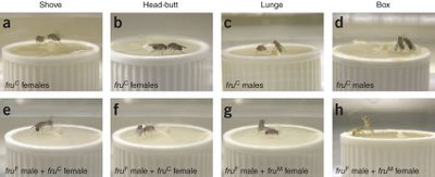
Figure 2. Aggresion Behavior in flies

When the versions of Fruitless are swapped, the males fight like females (the sissies) and females lunge at their opponents as seen in the Figure 2 above. The top panel shows the normal aggresion patterns seen in males and females while the bottom panel shows what happens when the flies produce the wrong version of the protein. Panels e and f show males exhibiting female aggression pattern when they express the female version of the protein. When the sexes with the opppsite version of the protein are put together in the panel g and h. In panel g, the upright lunging fly is a female and so is the upright "boxing" fly in panel h , indicating that swapping the protien alters how the flies respond - another innate behaviour affected!

The question that still remains (the most important one) is what is the effector? What does Fruitless, a transcription factor, modulate in a gender specific manner to control the sex specific aspect of behaviour?

There is a lot to still uncover but we are finally beginning to glimpse at how a genes influence how we respond. I believe that most behaviour is hard wired but at the same time modulation of the behaviour is environmental dependant. And now we finally are beginning to tell the effect of nature on nurture. A fun time lies ahead in molecular neuroscience!


Reference

1 - Demir, E. & Dickson, B. J. Cell 125, 785−794 (2005).

2- Vrontou E, Nilsen, S. P., Demir, E., Kravitz, E. A. & Dickson, B. J. Nature Neuroscience - 9, 1469 - 1471 (2006)

Sunday, November 19, 2006

Exploiting the parasites to our advantage

In a recent paper (Oct 20, 2006) in the journal Science, scientists have reported the use of a parasite-specific machinery in to correct certain deficiencies in human cells, which can be then used to tackle critical genetic disorders in humans.

This is cool for several reasons: taking lessons from a one-celled parasite to apply and solve complex genetic disorders in humans is cool by itself. What made me more happy is that the paper is from a group of scientists at the Indian Institute of Chemical Biology, Calcutta, India. One of the few (AFAIK, one of the first this year) all-indian authored papers from an Indian lab in the very prestigious journal Science. (Atleast in the field of molecular biology/diseases).

Eukaryotic cells are divided into compartments: the nucleus of the cell contains the genetic blueprint of the organism (instructions coded in DNA) and is responsible for it's maintenance, expression and regulation amongst other functions. The mitochondrion, known as the "power-house" of the cell, is where energy is produced from macromolecules through a series of biochemical reactions. The mitochondria possess their own set of genetic instructions (mtDNA) which store some mitochondria-specific instructions. Mutations in the mtDNA lead to some serious disorders given that the mitochondria are the energy-centres of the cell. One such syndrome is the Kearns-Sayre syndrome (KSS) : a nervous system disorder characterized in humans by hearing loss, difficulty in swallowing, loss of muscle co-ordination and cardiac function. KSS is caused by a large deletion in the mitochondrial DNA which disrupts mitochondrial function due to loss of the information encoded in that portion.

Fixing defects in mtDNA is challenging. The mitochondria are a double-membrane enclosed compartment; delivery of material to reach the mitochondrion and get incorporated is not trivial.

This is where scientists decided to take a leaf out of the book of the protist parasite Leishmania. Leishmania is a Trypanosomatid protist parasite that is transmitted by some species of the sandfly and causes leishmaniasis ( kala azar) that is endemic to several tropical and sub-tropical countries. This group of critters are characterized by some very divergent pathways in terms of their genetic organisation. One of these includes the fact that their mitochondrial genomes do not encode any tRNA genes. (tRNA refers to a set of genes involved in the making of proteins). As a result, the entire set of tRNA genes needs to be imported from the cytosol, for which these parasites have evolved a highly specialized machinery. The transport of tRNAs from the nucleus to the mitochondria is brought about by the RIC complex (RNA Import Complex) : a mulit-subunit complex found in the inner membrane of the leishmania mitochondria. The first significant accomplishment by Adhya and colleagues was to isolate this complex and purify it. Next. they show that Leishmania RIC is capable of transporting human tRNA molecules. Then, in a series of elegant experiments described in the science paper, they went on to test if human cells are capable of taking up leishmania RIC and using it to transport tRNA molecules to the mitochondria. They incubated a variety of human cell-cultures (including cells from patients of mitochondrial disorders) with Leishmania RIC and showed that RIC was successfully localized to the mitochondria of humans. Furthermore, they showed that the defective human cells were capable of transporting a specific tRNA molecule after incorporating the RIC, which they were unable to do earlier. Thus, RIC was capable of restoring defective human cells with the required mitochondrial function, showing great promise to reverse the effects of the inherited disorder. This has great therapeutic value in correcting several defects caused by various mitochondrial mutations which are hard to reach and correct otherwise.

Think about the myriad of distances travelled here: they have crossed millions of years on an evolutionary time scale to bring a mechanism from a one-celled critter to a multicellular human system. Then, at the cellular level, crossing multiple membranes and barriers to transport molecules to the right destination. Simply fascinating!

Ref: Mahata et al, Science 20 Oct 2006 vol. 314, pp 471-474.

Sunday, November 12, 2006

Biological control systems: (attempts in) Understanding the Nature's way

For anyone who has watched the evening twilight, dark clouds and the flock of wild geese fly across the sky, it is not difficult to figure out the relative motion among the birds and among the clouds...even from a moving vehicle. This may seem to be such a simple everyday experience that few really think twice about it, but this is considered one of the toughest problems in image processing. Posed in a more scientific terms, the problem is how can you distinguish the relative motion between two frames in a noisy environment? Any one who has fiddled with an SLR knows how difficult it is to get a perfect picture under varying environmental conditions. A slight tremor in hand can ruin a picture. However, our eyes do it without much concious effort in part due to the excellent in built control system that exactly regulates the amount of light entering the eye and focuses the image on retina. Other aspect of the built in control system is that it corrects for the movement of the head with an interface to the vestibular organ in the ear. Such fascinating control systems are part of every biological system governing almost every aspect of life...Respiration control, blood pressure and thermo regulation, circadian rhytms, chemical reaction in cells and many more. In this article we will discuss about the light regulation system in eye and a little bit about thermo regulation.
Let there be light:
As we think about the basic parts of our eyes (Fig. 1), we observe that they consist of cornea, lens, iris, ciliary muscle, and retina connected by optic nerve to brain. Functioning of eye depends on the amount of light entering the eye (Light intensity) and focusing of the image on retina. Two independent systems control these two aspects of vision.
Firstly, amount of light is regulated by the opening of pupil which is controlled by the two muscle groups in iris. Sphincter (controlled by cranial circuit which also controls the ciliary muscles which help in focusing) and dilator muscles (controlled by symphathetic nervous system of spinal cord: I wonder whether this is the reason why people look for dilated pupils when looking for vital signs in an unconcious person). Dilator muscles causes the pupil to open more whereas sphincter muscles cause the pupil to close as they contract. Acting together they control the opening of pupil in such a way that there is always an optimum light intensity falling over retina. This is a feedback control system where the pupil opening is the controlled variable( via the sphincter and dilatory muscles) while the system output is the light intensity on retina. The input is the light intensity of the environment and the control system aims to achieve a perfect pupil opening that optimizes the light intensity on retina under varying environmental conditions. By testing the pupil opening with narrow light beams (so that they do not have an effect on the light intensity regardless of pupil opening and thus disconnecting this feedback loop) it was found that the system is a very stable low gain system. Further, it is to be noted that this is not the only way eye responds to light intensity. There is another system on retina itself which adjust the signals to the optic fiber based on light intensity (reason why we can see the outlines a little better in a dark room after a few seconds of adaptation).

For more information about eye: http://www.arn.org/docs/glicksman/eyw_041001.htm

How about a little warmth as well: Maintianing optimum body temperature is vital for survival as most of the enzyme catalyzed reactions rates depend critically on it. Temperatures of cold blooded animals follows that of its surroundings (poikilothermy; one of the reasons such animals can be found mostly in tropical and temperate regions of the world) while that of warm blooded animals is tightly regulated (of course allowing for diurnal variations based on circadian rhytms) and is known as homeothermy. However, not everything is in pure black and white as is the rule in nature. During hibernation warm blooded animals such as hedgehog, bat and dormouse become coldblooded (to conserve their energy?) and this is referred to as heterothermy.
(More info on mammalian temperature regulation here: http://animals.about.com/cs/mammals/a/aa061601a.htm and Wikipedia entry:
http://en.wikipedia.org/wiki/Body_temperature )


It can be observed that for the thermoregulation system: the controlled variable is the heat producing/conserving mechanism while the output is body temperature. The input variable is the environment temperature and the temperature of the body. Now one can ask, where should be temperature be measured so that it best represents the body temperature? on the surface of the body? or closer to the internal organs? As anyone would point out, skin temperature is not the best place to estimate the body temperature, just as placing the thermometers on the outside of a building whose interior temperature has to adjusted is a bad idea. However, it is always a good idea to open/close the windows based on outside temperature while firing up the heater must be based on both outside and inside temperature. Something similar happens in our bodies too....the internal mechanism activates the heat producing/ conserving mechanism; while the case of conserving heat by closing windows regardless of inside temperature can be related to closure of sweat glands in cold weather regardless of internal temperature. This way the amount of energy expended to maintain the temperature can also be minimized. more often than not, such multiple optimization schemes are inbuilt in biological control systems.
Ref: Optimality principles in biology: Robert Rosen, Butterworths, London. 1967. [An Excellent book that deals with the issues we discussed in chapter 9]

In the next article, we hope to discuss how seemingly extremely complex branching pattern of blood circulation system can be derived from optimality arguments (again the above book has an excellent analysis).


The LISA Project

The Theory of Relativity has some rather fantastic claims to make about the nature of the universe. We still find it hard to believe, for example, that time slows down relative to the rest of the universe when you travel at speeds comparable to the speed of light. Imagine then, how the scientific community at the beginning of the 20th century felt.

In 1919, British astrophysicist Arthur Eddington conducted an experiment that confirmed one of the fundamental premises of Relativity. During a solar eclipse, they measured the angle of stars behind the sun... and found that the gravitational field of the Sun did in fact bend light passing near it. From then till now, scientists have been laying out the multifarious consequences of Einstein's theory and finding ways of physically confirming them. One of these consequences is the presence of "Gravitational Waves". To the layman, one would explain it as a "wobble" in the position and shape of all matter in the universe, arising from propagating distortions caused by massive gravitational interactions like the merging of supermassive black holes or a star spiralling into a black hole.

Figure 1: The effect of gravitational waves of the two different polarizations they can exhibit: "Plus" and "Cross" (Source: Chakrabarthy, 1999)

While there do exist projects to measure the effect of gravitational waves from Earth, the quality of the data is poor. Instead of taking measurements from celestial objects, scientists want to make more direct measurements. Enter the Laser Interferometer Space Antenna, or LISA project, scheduled to be launched in the year 2015. LISA consists of three spacecraft positioned at the vertices of an equilateral triangle. Using interferometry, they can measure minute variations in their relative positions, in the range of picometers!


Figure 2: Artist's rendering of LISA (Source: NASA)

Unfortunately, such variations can be caused by a variety of factors such as solar wind, gravitational field of nearby bodies, accumulation of electrostatic charges, etc. It is an engineering challenge to develop active and passive techniques to minimize these variations in order to get an accurate measurement relating only to gravitational waves. Until the spacecraft are actually operational, it is difficult to predict how successful we will be at this.

The presence of gravitational waves is more or less accepted scientific fact, and if the LISA project is successful, it will only be the final confirmation in this regard. The purpose of LISA is much more than just confirming the nature of gravitational waves. Since gravitational waves pass through all matter whereas light and other electromagnetic variation can be blocked by various "opaque" objects, accurate measurement of gravitational waves can yield information about parts of the universe currently inaccessible to us. We would be able to settle the issue of "dark matter". We would be able to get information on the nature of the universe much closer to the time of the Big Bang than we can now. Astrophysicists are lining up issues that the measurement of gravitational waves can settle.

Since the launch of LISA is nearly a decade away, for now we can just sit back and contemplate our wobbly natures.

References:

Indrajit Chakrabarthy, "Gravitational Waves: An Introduction," arXiv:physics/9908041 v1, Aug 21, 1999

LISA Project Home Page

Wikipedia on Gravitational Waves

Sunday, November 05, 2006

What is Life?

As one takes an evening stroll, one can probably distinguish between all the living and non-living objects that one encounters. In spite of a classic book by Erwin Schrodinger, with a title that seems to ask one for a definition of life, published in 1943 [1], the scientific community has still not been able to come up with a single answer to this fundamental question that satisfies every scientist. Part of the reason for this ambiguity is because, to date, there remains a controversy over which objects should be considered as living beings [2]. For example, can a virus be considered as a living being?

But, first let's try to discuss some of the traits of living things:

1. Metabolism: A living being consumes energy from the surroundings by converting one form of energy to other forms of energy by a process called metabolism. Metabolism, the Greek word for change, designates all the chemical reactions carried out within a living organism.

2. Organization: The energy gained from metabolism helps organisms to remain far more organized than non-living things. Organization here refers to the fact that one can not reduce an organism into smaller independent parts. All living organisms are formed of the basic biological unit called the cell. Within each cell, there are membranes that divide the living world from the non-living world and within the membranes, the cellular constituents are organized hierarchically to form a live entity. All the molecular constituents within the cell serve a function. These molecules are organized into an integrative system and serve the activities of the cell as a whole. Some people even argue that keeping this organization going is the basic entity of life, and the minute an object is dead, this organization is lost. One can study independent parts (as molecular biology) or cells for that matter, but in reality, life as we know it, can not exist without being organized at various levels hierarchically.

3. Reproduction: Living things can reproduce on their own to produce new organisms of the same kind. The instructions to reproduce are also inherent within an organism and are inherited by each generation from their parents.

4. Evolution: Living things are able to evolve over time on their own according to their environment. They evolve due to the occasional errors that crop up while copying the instruction from one generation to another. These errors track changes in the environment and an organism that is better adapted to the environment survives. Darwin's central contention was that this adaptation stems from the interplay of random variation and natural selection. So, the history is as important as organization to understand the workings of the present day organism.

An object is traditionally considered to be living if it has all the above characteristics [3]. In addition, the definition is applied at a global level to a whole species and not to individual beings [4]. In other words, sterile organisms are also considered to be alive even though they may have lost the ability to reproduce.

Non-living things may have one or more of the above mentioned traits, but do not possess all the above mentioned characteristics. For example, a flame can use up energy and convert chemical energy to light and heat energy, using up energy in this process. However, it can not reproduce on it's own and neither does a flame evolve according to it's environment.

Viruses on the other hand are a little more difficult to distinguish. They can evolve and they can reproduce (albeit, inside another organism and not on their own), but they do not possess any metabolic capabilities, and hence, it may be argued, should be considered as not living. A small minority of the biologists have postulated that the abilities to reproduce and evolve are the only criteria for life, and that viruses should hence be considered alive.

Seeds also form an interesting example. Do we consider seeds as living or non living? Well, I did a google search and they are considered to be alive. They certainly have the ability to reproduce and, hence, evolve under the "right conditions". In addition, they are as organized as a living organism, but the real question was whether metabolism takes place in a seed under dry storage conditions. I was pleasantly surprised to find many papers reporting that seeds do undergo metabolism even during storage (an example is [5]), and hence, they do have all the criteria to be considered alive.

Physicists and chemists tend to argue over whether all the four properties are really required for life. While some chemists argue that metabolism is the real criterion for life, physicists argue that the level of organization in a cell is what really demarcates the difference between a living cell and a non-living cell. In fact, an algebraic information theoretic framework was developed to define the amount of information required to define an organism and the amount of organization in an organism [6].

What should be considered as living is not only an academic issue, but is equally important for space probes that look for signs of extraterrestrial life. In addition, it is equally important when one studies the origin of life from non-living entities. When does one consider that there is enough complexity in a system to call it a living cell? I will continue this post with a post on the quest for the origin of life and also, on a separate series of posts, on molecular evolution of living organisms.

References:
[1] What is Life? by Erwin Schrodinger.

[2] Chapters 1 and 2 of The Way of the Cell by Franklin Harold.

[3] Wikipedia entry on Life.

[4] Brittanica Encyclopedia.

[5] Metabolic activities of dormant seeds during dry storage. Naturwissenschaften, 59:3, 1972, 73-74.

[6] Toward a Mathematical Definition of "Life" by Gregory C Chaitin.

Thursday, November 02, 2006

In Living Color (Part 1)

This is the first in a series of posts that will describe how light and optical technologies are playing an important role in modern biological investigations. This post is about fluorescent proteins, their history and some applications.

It is often said that the 21st century will be (is) the age of biology; much like the previous century was for physics. New discoveries are occurring and biological information is growing both in size and complexity at an exponential rate. A major factor fueling this growth is the plethora of technologies available to the modern biologists in their quest to uncover the very basic molecular mechanisms of life.


Among such tools, the discovery and use of fluorescent (light emitting) proteins, has proved to be a major boon for scientists investigating activities of genes and proteins inside cells. Fluorescence is an optical phenomenon, where a molecule absorbs photons of a particular color and thereafter emits photons of a different color - with the emitted color always red-shifted. While this phenomenon had been observed in living organisms, the molecules involved in the luminescence were unknown till the 1960s. In ‘62, Osamu Shimomura, a Princeton scientist investigating the phenomenon of bioluminescence, isolated a light-emitting protein from the jellyfish, Aequorea victoria, in the Padget Sound area of Washington state. This protein, named aequorin1, produced blue light – but only in presence of calcium. However, as a footnote in the publication of this discovery, Shimomura and co-worker mentioned “….a protein giving solutions that look slightly greenish in sunlight through only yellowish under tungsten lights, and exhibiting a very bright, greenish fluorescence in the ultraviolet of a Mineralite, has also been isolated…”. It was soon found that this ‘other protein’ was involved in absorbing the blue light from Aequerin and emitting the green light observed in the jellyfish. It was named appropriately, even if perhaps a little unimaginatively, the green fluorescent protein (GFP).


The jellyfish, Aequorea victoria, on the left and green bioluminescence observed around the margin (note the picture on the left does not show fluorescence !)


However, it took another thirty years before the GFP became the almost ubiquitous cellular and molecular biology tool it is today. In 1987, Doug Prasher, then at the Woods Hole Oceanographic Institute, discovered and was able to make a copy of the DNA sequence within the jellyfish gene that encoded for GFP. He did not, however, succeed in making a glowing protein from the DNA sequence in the lab. Subsequently, Prasher sent his sequence to a researcher at Columbia, Marty Chalfie who was able to produce the protein in bacteria - a typical trick used by biologists to make proteins. As shown in the figure on the right from Chalfie's work, the bacteria containing the genes for GFP (on the right side of the plate) emits green light under illumination with ultaviolet lamp (it was a graduate student doing rotation in Chalfie’s lab that actually performed the work and made the discovery!). This seminal work, published in 1994 in the journal Science, led to 'an explosion of color' in the biological world. Subsequently, other researchers showed that the GFP could be produced, alone or in tandem with other proteins in a variety of organisms.

Over the last decade, a great deal of research has contributed towards understanding the underlying physical mechanisms of GFP’s light emission2 and importantly, towards improving its properties through genetic manipulation. The leader in this field has been Roger Tsien, who along with co-workers demonstrated that making small changes, such as replacing a few amino acids in GFP could make it glow brighter, mature faster and prevent aggregation of the protein inside cells. His group has also succeeded in tuning the absorption and emission of the original GFP through mutagenesis, leading to a veritable palette of fluorescent proteins that absorb and emit light through the entire span of the visible light spectrum (see below). Additionally, a Russian scientist, Sergey Lukyanov, used the GFP sequence as a 'bait' to search for novel fluorescent proteins in corals and succeeded in finding several GFP-like proteins, particularly a red-emitting fluorescent protein, dsRED from Anthozoa, which is also used widely.


Panel on top shows the fluorescent protein 'palette' developed by Tsien lab - note range of colors and the fruity names. On the left, artwork with bacteria expressing various colors of fluorescent protein.


The major advantage of GFP is that inside a living cell, it can emit light on its own without the help of another protein or other chemicals. It is also possible by using molecular biology techniques, to attach the DNA of GFP to the DNA of the protein of your choice to produce a recombinant DNA. When the information from such recombinant DNA gets translated into a protein within the cell, a tandem protein is created with the GFP unit hanging from the protein. Importantly, since the size of GFP is relatively small, in most cases it does not interfere with the regular functions of the protein it is attached to.

In the simplest of applications, after shining light on the cells, the total amount of fluorescence obtained from the cells provides a measure of the level of expression of the protein tagged with GFP. However the more useful applications involve cells placed under microscopes with high magnifying power (40x to 100x) in conjunction with either arc lamps or lasers for bright illumination and high-resolution detection devices such as CCD cameras for
capturing images of the emitted light. In these cases, we can literally see where the protein of interest is located, or illuminate a particular subcellular structure. For example, the figure on the right shows the mesh of protein network that act as a 'skeleton' (in fact it is called the 'cytoskeleton') in majority of cells in higher organisms. A protein called 'actin' that is involved in this scaffold has been tagged with GFP.

It is also possible to tag two or more proteins in the cells with different fluorescent colors (see the fluorescent protein palette above) and follow their localization or movement in cells. This helps in noting where two proteins are localized during a cellular function. In the figure to the right, the protein actin is now tagged with a cyan emitting fluorescent protein (CFP). Another protein, vinculin, has been tagged with a yellow fluorescent protein (YFP). You can observe that the YFPs are localized as small elliptical structures at many places. These are called focal adhesions, which form a link between the cell cytoskeleton (in cyan) and its extra-cellular matrix. This interaction help cells to adhere and eventually move about in a tissue.

(in a future post, I will talk about a technique involving fluorescent proteins of two colors which is used to determine if two proteins interact with each other inside a cell)

Perhaps the most powerful application of fluorescent proteins is when you combine microscopy with time lapse video images. In such cases, it is possible to observe where and when the translocation of the protein in cells is taking place under a biological condition. For example, see a video here of a cell moving around with a protein involved in the focal adhesion tagged with GFP.

Visualization of protein location and dynamics in this manner enable scientists to place cells under various physiological conditions and observe the resultant phenotype of the protein behavior. Before the advent of GFP, scientist had to destroy the cells and use other tedious biochemical techniques to obtain similar information. Even then real-time data acquisition was not possible.

A quick search of the database will reveal more than ten of thousands of peer-reviewed publications where fluorescent proteins have been used to study protein functions at the cellular level. In most of these cases, research was conducted with either unicellular organisms or cells derived from tissues of mammals. However, apart from single cells, fluorescent proteins are also being used at the tissue and even the whole organism level. The picture below shows an example of a research which is investigating the movement of neurons (labeled with GFP) in the cerebral cortex.


A more well-known example of GFP in whole organisms, is the development of 'fluorescent mice' by the company Anticancer Inc . It is easy to follow tumor progression and cancer metastasis in such mice. Also, a Taiwanse reasearch group recently created 'fluorescent pigs'. Stem cells or organs from these pigs when transplanted into other organisms can be followed easily without requiring invasive techniques.


Animal Farm: fluorescent pigs and mice.


More examples of such applications of GFP can be found here. Apart from these animals, 'Alba' , the fluorescent rabbit and fluorescent aquarium fishes are two examples of more esoteric application of this scientific technology.

On a final note, betting markets for the Nobel Prize (yes they do exist !), were predicting this year’s Chemistry Nobel to go to Roger Tsein and others for their work on fluorescent proteins. It eventually went to Roger Kornberg for his work on DNA transcription. Considering the importance of fluorescence proteins and their wide-ranging revolutionary impact on biology, it is not far-fetched to think that the Nobel is not beyond the grasp of these researchers.

Notes:

1. Aequorin itself has been very useful for visualizing cellular calcium concentrations, the regulation of which is important for a number of physiological activities.

2. Without going into great details about physi-chemical mechanisms of GFP fluorescence, suffice to say that the protein has a barrel-like structure (see below); within the barrel, three critical amino acids are brought together in close spatial proximity, which forms the chromophore.


Artistic rendition of the three-dimensional structure of GFP.


3. All images have been linked back to their original web-page.

4. Recommended further reading: This web-site is a very good resource for learning more about GFP's discovery, structure and applications. Also read this interview with Dr. Martin Chalfie.


Coming up: "Much to fret about ": on a technique known as fluorescence resonance energy transfer that enables biological distance measurements, detection of protein interactions, and can be used to look at protein functions at a single molecule level !