Many people have wrote that there is no such thing as altruism. I must admit that I have also always thought the same thing..even though I continued to search for one such act. I have often been the lone arguer pointing out that no one has been able to uncover a single example of true altruism...in a natural setting or in the human race. I am now rescinding my previous statement. I have for a very long time..searched for an example that I could in some way attribute to altruism..and until now I have been unsuccessful. I suppose all examples are based upon your own definition of altruism. I learned many years ago that altruism is an act done by an individual at a cost..benefiting another individual not of relation to the one performing the act.
The said example concerns a chimpanzee and a human. A researcher was following a group of chimpanzees in the jungle. After some hours, he found that he had forgotten his lunch back at the research station. This researcher then proceeded to try to knock down fruit from a tree some distance from where the group of chimpanzees sat eating their mid-day meal. It has been noted that after some time of unsuccessful attempts to acquire fruit... a young male from the group collected some fruits from a tree and climbed down toward the researcher. The chimpanzee then proceeded to approach the researcher and leave the fruit for the researcher.
This instance has been noted as a true act of altruism by any definition since the chimpanzee was not of relation to the researcher (not in the last 1000 years at least) and this act was a cost to itself with no benefit. Therefore, I stand corrected on the notion of altruism...it seems to exist after all.
References
Compassion, Rescue and the Altruism Debate, The emotional lives of animals Jeffrey Moussaieff Masson and Susan McCarthy.
Saturday, December 23, 2006
Tuesday, December 19, 2006
Why We Should Always Question..

There are many moments in time that we look back at now...which will always lead to us to slap our foreheads and say, "stupid, stupid, stupid". There is one such moment that continues to be brought up in the most general science classes..as if to say.."if you don't test and retest...we will ridicule you for years to come". This example is of course the lemming. Lemmings were believed to commit, "The Lemming Suicide Plunge" when the population became too numerous. It was really believed that millions of lemmings would be overcome by a hard-wired impulse to dash to their death by hurling themselves over a cliff to the rocks below or by plunging into the sea to die a horrible death by drowning. The reasoning behind this was thought to be a deep-rooted act of altruistic behavior resulting the greater good for the species. However, you can probably guess, this is not the case. Shown to the right is a famous Far Side cartoon dipicting natural selection in action. As the lemmings dash to their death..there is one cheater in the group that will survive and ultimately passing on the cheater genes to future generations.
How this story may have gotten started....
Many rodent species experience very strange cyclic population explosions. It is very interesting that lemmings have one of the most regular cyclic fluctuations in population densities. It has been shown that these little creatures have population explosions about every three or four years. The population numbers of lemmings explode to high numbers, and then drop almost to extinction. Even after approximately 75 years of intense research, scientists do not fully understand why the populations fluctuate so much. Throughout the years, many factors have been tested (i.e., changes in food availability, climate, density of predators, stress of overcrowding, infectious diseases, snow conditions, sunspots, etc) but none completely explain why populations of lemmings have these explosive cycles.
The myth of the lemming most likely started when these population explosions happen and the lemmings migrate away from areas with a dense population. As you can imagine, the migrations begin slowly and erratically. It has been shown that small numbers of lemmings will move at night, and larger groups in the daytime. This movement causes small groupings of lemmings to move instead of one continous mass, usually seperated by a short time frame of 10 minutes or so. It has been noted that they will often follow well worn paths and roads along their journey.
As you can imagine, there will be unavoidable obstacles, such as streams and lakes inevitalby causing them to swim as a last resort. Suprisingly, they are able to swim across a 200 meter body of water on a calm night, but most will drown in a windy night.
So why the myth began....
It is due to a very unlikely source....Walt Disney. Walt Disney was making a movie tittled, "Wild Wilderness" which was released in 1958. It was filmed in Alberta, Canada, in a location that is far from the sea and not a native home to lemmings. The lemming were imported and forced to jump to thier deaths by placing them on a spinning turntable that was covered with snow, and then shooting it from many different angles. The cliff-death-plunge sequence was done by herding the lemmings over a small cliff into a river. It's easy to understand why the filmmakers did this - wild animals are notoriously uncooperative, and a migration-of-doom followed by a cliff-of-death sequence is far more dramatic to show than the lemmings' self-implemented population-density management plan.
The moral of the story....lemmings do not commit mass suicide and Walt Disney has clear prejudice against the lemming and for the mouse.
Wednesday, December 13, 2006
Net Picks
Something to read while taking a break -
Fifty Years With Double Stranded RNA
Alexander Rich, the scientist who discovered hybridization and the "other" double helix describes what it meant to biology.
Bioengineering and AIDS
University of Utah scientists designed a "molecular condom" women could use daily to prevent AIDS by vaginally inserting a liquid that would turn into a gel-like coating and then, when exposed to semen, return to liquid form and release an antiviral drug.
Genetic Map Offers New Tool For Malaria Research
In one of three genomic studies of malaria appearing in Nature Genetics, scientists chart genetic variation across the genome of the malaria parasite, unlocking novel DNA regions associated with drug resistance.
Laugh And The Whole World Laughs With You: Why The Brain Just Can't Help Itself
Researchers at UCL (University College London) and Imperial College London have shown that positive sounds trigger a response in the listener's brain in an area that is activated by smile, as though preparing our facial muscles to laugh.
This Holiday Season drink without fear.
A study performed by the Research Laboratories of the Catholic University of Campobasso (Italy) confirms the beneficial effects that moderate consumption of alcohol has on our health: drinking in moderation reduces all-cause mortality.
Another reason why the Octopus Rules - in built 3-D light reflectors!
Roger Hanlon at the Marine Biological Laboratory, Woods Hole, Massachusetts and colleagues took a close look at the octopus's skin and identified a new group of proteins (leucophores) with remarkable properties.
New Insights Into The Origin Of Life On Earth
In an advance toward understanding the origin of life on Earth, scientists have shown that parts of the Krebs cycle can run in reverse, producing biomolecules that could jump-start life with only sunlight and a mineral present in the primordial oceans.
Wednesday, December 06, 2006
Science Daily Picks
Found : Apple Gene for Red
CSIRO scientist have found the gene that controls the color of apples. The red color of apple skin is result of anthocyanins (responsible for blue or red color in plants). It was known that color of apples is affected by light (less light results in poor color), so the scientist looked for genes that were activated by light and compared these to genes in a green apple and bingo!
Learning During Sleep?
Max Planck Institute for Medical Research in Heidelberg have taken a step towards understanding communication between memory areas. Their study shows a link between sleep and memory consolidation (transfer of memory from hippocampus to the cerebral cortex).
Cities Change The Songs Of Birds Leiden University researchers studied the songs of the great tit (Parus major), a successful urban dweller in the center of ten major European cities and compared the songs to that of the bird living in nearby forest sites. The results show that songs that are important for mate selection or territory defense are shorter, sung faster and at a higher pitch by the urban bird. The findings published on Dec.5 in Current Biology show divergence within a species because of the environment and could very well lead to speciation.
NASA Telescope Sees Black Hole Munch On A Star.
NASA’s Galaxy Evolution Explorer has caught a black hole eating a star, from the initial capture to the last bites.
Water Still Flows In Brief Spurts On Mars, NASA Images Suggest
NASA photographs have revealed bright new deposits seen in two gullies on Mars that suggest water carried sediment through them sometime during the past seven years.
So let the hunt for the ETs begin :)
Monday, November 20, 2006
Fruitless Attempts To Explain Behaviour Hardwiring
About an year ago, Dickson lab had published an article about a behavioral switch gene, called Fruitless. It had gained notoriety in the popular press as the “Gay Gene” since it affects sexual orientation but to me Fruitless is interesting not only because it determines sexual orientation but more importantly as it gives a glimpse into how genes modulate behavior.
You see, behavior is “hard wired”into our nervous system during development. The neuronal body plan is already present in our genome - the developmental genes that direct cells to grow into a specific type or in a specific direction. These genes during development take cues from the environment and hard wire innate behavior in to a species. I have always thought that to be the coolest thing. Think about it - we are not only a function of our genes but also product of our environment. And Dickson's research links the two together - how a presence of a single gene product directs the function of neurons responsible for sexual orientation in fruit fly males. Courtship behaviour in D. melanogaster is invovles a series of well chorographed steps that invovle the visual, the olfactory, the tactile, the acoustic, the gustatory and the mechanosensory stimuli being exchanged between the sexes (See Fig.1). The role of the female is more simplified -she simply runs away, gives the odd kick, then mates (or not).
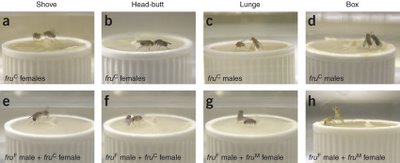 Figure 2. Aggresion Behavior in flies
You see, behavior is “hard wired”into our nervous system during development. The neuronal body plan is already present in our genome - the developmental genes that direct cells to grow into a specific type or in a specific direction. These genes during development take cues from the environment and hard wire innate behavior in to a species. I have always thought that to be the coolest thing. Think about it - we are not only a function of our genes but also product of our environment. And Dickson's research links the two together - how a presence of a single gene product directs the function of neurons responsible for sexual orientation in fruit fly males. Courtship behaviour in D. melanogaster is invovles a series of well chorographed steps that invovle the visual, the olfactory, the tactile, the acoustic, the gustatory and the mechanosensory stimuli being exchanged between the sexes (See Fig.1). The role of the female is more simplified -she simply runs away, gives the odd kick, then mates (or not).

Fig. 1 Courtship behaviour in Fruitfly
This normal courtship behaviour seems to be disrupted in the Fruitess mutants. Before we talk about what happens when we mutate this gene, lets us take a brief overview of what is known about the gene. Fruitless gene was molecularly cloned in 1996 and the putative protein encodes a transcription factor. Fruitless is sex specifically spliced - in lay man terms it means that males produce one version of this protein where as the females produce another. This sex specific splicing is regulated by presence or absence of another protein called Transformer, which in Drosophila also determines the sex in the fly.
So what happens if you produce the wrong version of the protein in either sexes? By forcing males to express the female-specific Fruitless transcript by using the awesome power of Fly genetics (:P), the Dickson Lab produced males that were sterile, uninterested in courting females, actively courting males, actually ending up forming courtship chains (see this). By contrast, females making the male version of the protein mated poorly, produced very few eggs, but — astonishingly — courted other females (see this), even to the point of forming chains.
So why does this happen? When you look at the the central nervous system of males and females ,there are very few differences in terms of sex-specific Fruitless expression- in number, position or wiring of cells that express this protein. But Fruitless is present in the olfactory sensory neurons which play an important role in fly courtship behavior. So when male fruit flies cannot produce this male specific form or produce a mutant form of this protein, you get males that court other males. In other words, a single gene encoded product is enought to shift the functioning of the nervous system from male to female mode, irrespective of the morphological sex of the animal. Simply put with mutant (rather non-sex specific)versions of the protein, flies change their sexual orientation but they not other aspects of their morphology.
Now the same gene is making a news splash again – the Kravitz lab has linked Fruitless to yet another sex-specific behavior – aggression/ fighting patterns. Aggression found in almost all animals - from sea anemones to human - helps to acquire food/shelter/ mates or defend the same. Despite its importance, relatively little is known of the neural and humoral mechanisms that are its proximate causes. Many behavioral patterns in aggressive behavior are shared in flies but there are a subset that are sex specific. Female fighting, for example, largely involves head butts and some shoving. Males show extended wing threats, wing-flicking while retreating, and high intensity components of fighting like boxing, tussling and holding. In contrast to male fighting behavior, no clear hierarchical relationship results from the interactions between female flies.
 Figure 2. Aggresion Behavior in flies
When the versions of Fruitless are swapped, the males fight like females (the sissies) and females lunge at their opponents as seen in the Figure 2 above. The top panel shows the normal aggresion patterns seen in males and females while the bottom panel shows what happens when the flies produce the wrong version of the protein. Panels e and f show males exhibiting female aggression pattern when they express the female version of the protein. When the sexes with the opppsite version of the protein are put together in the panel g and h. In panel g, the upright lunging fly is a female and so is the upright "boxing" fly in panel h , indicating that swapping the protien alters how the flies respond - another innate behaviour affected!
The question that still remains (the most important one) is what is the effector? What does Fruitless, a transcription factor, modulate in a gender specific manner to control the sex specific aspect of behaviour?
There is a lot to still uncover but we are finally beginning to glimpse at how a genes influence how we respond. I believe that most behaviour is hard wired but at the same time modulation of the behaviour is environmental dependant. And now we finally are beginning to tell the effect of nature on nurture. A fun time lies ahead in molecular neuroscience!
Reference
1 - Demir, E. & Dickson, B. J. Cell 125, 785−794 (2005).
2- Vrontou E, Nilsen, S. P., Demir, E., Kravitz, E. A. & Dickson, B. J. Nature Neuroscience - 9, 1469 - 1471 (2006)
Sunday, November 19, 2006
Exploiting the parasites to our advantage
In a recent paper (Oct 20, 2006) in the journal Science, scientists have reported the use of a parasite-specific machinery in to correct certain deficiencies in human cells, which can be then used to tackle critical genetic disorders in humans.
This is cool for several reasons: taking lessons from a one-celled parasite to apply and solve complex genetic disorders in humans is cool by itself. What made me more happy is that the paper is from a group of scientists at the Indian Institute of Chemical Biology, Calcutta, India. One of the few (AFAIK, one of the first this year) all-indian authored papers from an Indian lab in the very prestigious journal Science. (Atleast in the field of molecular biology/diseases).
Eukaryotic cells are divided into compartments: the nucleus of the cell contains the genetic blueprint of the organism (instructions coded in DNA) and is responsible for it's maintenance, expression and regulation amongst other functions. The mitochondrion, known as the "power-house" of the cell, is where energy is produced from macromolecules through a series of biochemical reactions. The mitochondria possess their own set of genetic instructions (mtDNA) which store some mitochondria-specific instructions. Mutations in the mtDNA lead to some serious disorders given that the mitochondria are the energy-centres of the cell. One such syndrome is the Kearns-Sayre syndrome (KSS) : a nervous system disorder characterized in humans by hearing loss, difficulty in swallowing, loss of muscle co-ordination and cardiac function. KSS is caused by a large deletion in the mitochondrial DNA which disrupts mitochondrial function due to loss of the information encoded in that portion.
Fixing defects in mtDNA is challenging. The mitochondria are a double-membrane enclosed compartment; delivery of material to reach the mitochondrion and get incorporated is not trivial.
This is where scientists decided to take a leaf out of the book of the protist parasite Leishmania. Leishmania is a Trypanosomatid protist parasite that is transmitted by some species of the sandfly and causes leishmaniasis ( kala azar) that is endemic to several tropical and sub-tropical countries. This group of critters are characterized by some very divergent pathways in terms of their genetic organisation. One of these includes the fact that their mitochondrial genomes do not encode any tRNA genes. (tRNA refers to a set of genes involved in the making of proteins). As a result, the entire set of tRNA genes needs to be imported from the cytosol, for which these parasites have evolved a highly specialized machinery. The transport of tRNAs from the nucleus to the mitochondria is brought about by the RIC complex (RNA Import Complex) : a mulit-subunit complex found in the inner membrane of the leishmania mitochondria. The first significant accomplishment by Adhya and colleagues was to isolate this complex and purify it. Next. they show that Leishmania RIC is capable of transporting human tRNA molecules. Then, in a series of elegant experiments described in the science paper, they went on to test if human cells are capable of taking up leishmania RIC and using it to transport tRNA molecules to the mitochondria. They incubated a variety of human cell-cultures (including cells from patients of mitochondrial disorders) with Leishmania RIC and showed that RIC was successfully localized to the mitochondria of humans. Furthermore, they showed that the defective human cells were capable of transporting a specific tRNA molecule after incorporating the RIC, which they were unable to do earlier. Thus, RIC was capable of restoring defective human cells with the required mitochondrial function, showing great promise to reverse the effects of the inherited disorder. This has great therapeutic value in correcting several defects caused by various mitochondrial mutations which are hard to reach and correct otherwise.
Think about the myriad of distances travelled here: they have crossed millions of years on an evolutionary time scale to bring a mechanism from a one-celled critter to a multicellular human system. Then, at the cellular level, crossing multiple membranes and barriers to transport molecules to the right destination. Simply fascinating!
Ref: Mahata et al, Science 20 Oct 2006 vol. 314, pp 471-474.
Sunday, November 12, 2006
Biological control systems: (attempts in) Understanding the Nature's way
For anyone who has watched the evening twilight, dark clouds and the flock of wild geese fly across the sky, it is not difficult to figure out the relative motion among the birds and among the clouds...even from a moving vehicle. This may seem to be such a simple everyday experience that few really think twice about it, but this is considered one of the toughest problems in image processing. Posed in a more scientific terms, the problem is how can you distinguish the relative motion between two frames in a noisy environment? Any one who has fiddled with an SLR knows how difficult it is to get a perfect picture under varying environmental conditions. A slight tremor in hand can ruin a picture. However, our eyes do it without much concious effort in part due to the excellent in built control system that exactly regulates the amount of light entering the eye and focuses the image on retina. Other aspect of the built in control system is that it corrects for the movement of the head with an interface to the vestibular organ in the ear. Such fascinating control systems are part of every biological system governing almost every aspect of life...Respiration control, blood pressure and thermo regulation, circadian rhytms, chemical reaction in cells and many more. In this article we will discuss about the light regulation system in eye and a little bit about thermo regulation.
Let there be light:
As we think about the basic parts of our eyes (Fig. 1), we observe that they consist of cornea, lens, iris, ciliary muscle, and retina connected by optic nerve to brain. Functioning of eye depends on the amount of light entering the eye (Light intensity) and focusing of the image on retina. Two independent systems control these two aspects of vision.
Let there be light:
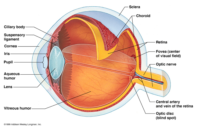
As we think about the basic parts of our eyes (Fig. 1), we observe that they consist of cornea, lens, iris, ciliary muscle, and retina connected by optic nerve to brain. Functioning of eye depends on the amount of light entering the eye (Light intensity) and focusing of the image on retina. Two independent systems control these two aspects of vision.
Firstly, amount of light is regulated by the opening of pupil which is controlled by the two muscle groups in iris. Sphincter (controlled by cranial circuit which also controls the ciliary muscles which help in focusing) and dilator muscles (controlled by symphathetic nervous system of spinal cord: I wonder whether this is the reason why people look for dilated pupils when looking for vital signs in an unconcious person). Dilator muscles causes the pupil to open more whereas sphincter muscles cause the pupil to close as they contract. Acting together they control the opening of pupil in such a way that there is always an optimum light intensity falling over retina. This is a feedback control system where the pupil opening is the controlled variable( via the sphincter and dilatory muscles) while the system output is the light intensity on retina. The input is the light intensity of the environment and the control system aims to achieve a perfect pupil opening that optimizes the light intensity on retina under varying environmental conditions. By testing the pupil opening with narrow light beams (so that they do not have an effect on the light intensity regardless of pupil opening and thus disconnecting this feedback loop) it was found that the system is a very stable low gain system. Further, it is to be noted that this is not the only way eye responds to light intensity. There is another system on retina itself which adjust the signals to the optic fiber based on light intensity (reason why we can see the outlines a little better in a dark room after a few seconds of adaptation).
For more information about eye: http://www.arn.org/docs/glicksman/eyw_041001.htm
How about a little warmth as well: Maintianing optimum body temperature is vital for survival as most of the enzyme catalyzed reactions rates depend critically on it. Temperatures of cold blooded animals follows that of its surroundings (poikilothermy; one of the reasons such animals can be found mostly in tropical and temperate regions of the world) while that of warm blooded animals is tightly regulated (of course allowing for diurnal variations based on circadian rhytms) and is known as homeothermy. However, not everything is in pure black and white as is the rule in nature. During hibernation warm blooded animals such as hedgehog, bat and dormouse become coldblooded (to conserve their energy?) and this is referred to as heterothermy.
(More info on mammalian temperature regulation here: http://animals.about.com/cs/mammals/a/aa061601a.htm and Wikipedia entry:
http://en.wikipedia.org/wiki/Body_temperature )
It can be observed that for the thermoregulation system: the controlled variable is the heat producing/conserving mechanism while the output is body temperature. The input variable is the environment temperature and the temperature of the body. Now one can ask, where should be temperature be measured so that it best represents the body temperature? on the surface of the body? or closer to the internal organs? As anyone would point out, skin temperature is not the best place to estimate the body temperature, just as placing the thermometers on the outside of a building whose interior temperature has to adjusted is a bad idea. However, it is always a good idea to open/close the windows based on outside temperature while firing up the heater must be based on both outside and inside temperature. Something similar happens in our bodies too....the internal mechanism activates the heat producing/ conserving mechanism; while the case of conserving heat by closing windows regardless of inside temperature can be related to closure of sweat glands in cold weather regardless of internal temperature. This way the amount of energy expended to maintain the temperature can also be minimized. more often than not, such multiple optimization schemes are inbuilt in biological control systems.
Ref: Optimality principles in biology: Robert Rosen, Butterworths, London. 1967. [An Excellent book that deals with the issues we discussed in chapter 9]
In the next article, we hope to discuss how seemingly extremely complex branching pattern of blood circulation system can be derived from optimality arguments (again the above book has an excellent analysis).
In the next article, we hope to discuss how seemingly extremely complex branching pattern of blood circulation system can be derived from optimality arguments (again the above book has an excellent analysis).
The LISA Project
The Theory of Relativity has some rather fantastic claims to make about the nature of the universe. We still find it hard to believe, for example, that time slows down relative to the rest of the universe when you travel at speeds comparable to the speed of light. Imagine then, how the scientific community at the beginning of the 20th century felt.
In 1919, British astrophysicist Arthur Eddington conducted an experiment that confirmed one of the fundamental premises of Relativity. During a solar eclipse, they measured the angle of stars behind the sun... and found that the gravitational field of the Sun did in fact bend light passing near it. From then till now, scientists have been laying out the multifarious consequences of Einstein's theory and finding ways of physically confirming them. One of these consequences is the presence of "Gravitational Waves". To the layman, one would explain it as a "wobble" in the position and shape of all matter in the universe, arising from propagating distortions caused by massive gravitational interactions like the merging of supermassive black holes or a star spiralling into a black hole.
 Figure 1: The effect of gravitational waves of the two different polarizations they can exhibit: "Plus" and "Cross" (Source: Chakrabarthy, 1999)
While there do exist projects to measure the effect of gravitational waves from Earth, the quality of the data is poor. Instead of taking measurements from celestial objects, scientists want to make more direct measurements. Enter the Laser Interferometer Space Antenna, or LISA project, scheduled to be launched in the year 2015. LISA consists of three spacecraft positioned at the vertices of an equilateral triangle. Using interferometry, they can measure minute variations in their relative positions, in the range of picometers!
 Figure 2: Artist's rendering of LISA (Source: NASA)
In 1919, British astrophysicist Arthur Eddington conducted an experiment that confirmed one of the fundamental premises of Relativity. During a solar eclipse, they measured the angle of stars behind the sun... and found that the gravitational field of the Sun did in fact bend light passing near it. From then till now, scientists have been laying out the multifarious consequences of Einstein's theory and finding ways of physically confirming them. One of these consequences is the presence of "Gravitational Waves". To the layman, one would explain it as a "wobble" in the position and shape of all matter in the universe, arising from propagating distortions caused by massive gravitational interactions like the merging of supermassive black holes or a star spiralling into a black hole.
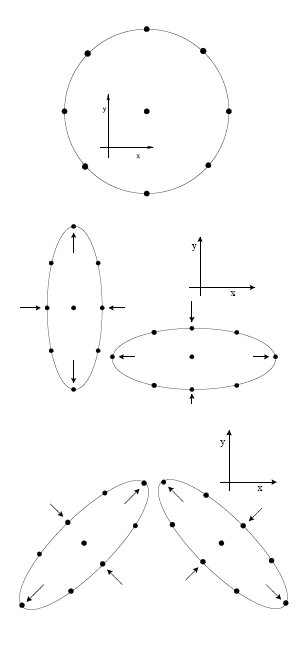 Figure 1: The effect of gravitational waves of the two different polarizations they can exhibit: "Plus" and "Cross" (Source: Chakrabarthy, 1999)
While there do exist projects to measure the effect of gravitational waves from Earth, the quality of the data is poor. Instead of taking measurements from celestial objects, scientists want to make more direct measurements. Enter the Laser Interferometer Space Antenna, or LISA project, scheduled to be launched in the year 2015. LISA consists of three spacecraft positioned at the vertices of an equilateral triangle. Using interferometry, they can measure minute variations in their relative positions, in the range of picometers!
 Figure 2: Artist's rendering of LISA (Source: NASA)
Unfortunately, such variations can be caused by a variety of factors such as solar wind, gravitational field of nearby bodies, accumulation of electrostatic charges, etc. It is an engineering challenge to develop active and passive techniques to minimize these variations in order to get an accurate measurement relating only to gravitational waves. Until the spacecraft are actually operational, it is difficult to predict how successful we will be at this.
The presence of gravitational waves is more or less accepted scientific fact, and if the LISA project is successful, it will only be the final confirmation in this regard. The purpose of LISA is much more than just confirming the nature of gravitational waves. Since gravitational waves pass through all matter whereas light and other electromagnetic variation can be blocked by various "opaque" objects, accurate measurement of gravitational waves can yield information about parts of the universe currently inaccessible to us. We would be able to settle the issue of "dark matter". We would be able to get information on the nature of the universe much closer to the time of the Big Bang than we can now. Astrophysicists are lining up issues that the measurement of gravitational waves can settle.
Since the launch of LISA is nearly a decade away, for now we can just sit back and contemplate our wobbly natures.
References:
Indrajit Chakrabarthy, "Gravitational Waves: An Introduction," arXiv:physics/9908041 v1, Aug 21, 1999
LISA Project Home Page
Wikipedia on Gravitational Waves
Sunday, November 05, 2006
What is Life?
As one takes an evening stroll, one can probably distinguish between all the living and non-living objects that one encounters. In spite of a classic book by Erwin Schrodinger, with a title that seems to ask one for a definition of life, published in 1943 [1], the scientific community has still not been able to come up with a single answer to this fundamental question that satisfies every scientist. Part of the reason for this ambiguity is because, to date, there remains a controversy over which objects should be considered as living beings [2]. For example, can a virus be considered as a living being?
But, first let's try to discuss some of the traits of living things:
1. Metabolism: A living being consumes energy from the surroundings by converting one form of energy to other forms of energy by a process called metabolism. Metabolism, the Greek word for change, designates all the chemical reactions carried out within a living organism.
2. Organization: The energy gained from metabolism helps organisms to remain far more organized than non-living things. Organization here refers to the fact that one can not reduce an organism into smaller independent parts. All living organisms are formed of the basic biological unit called the cell. Within each cell, there are membranes that divide the living world from the non-living world and within the membranes, the cellular constituents are organized hierarchically to form a live entity. All the molecular constituents within the cell serve a function. These molecules are organized into an integrative system and serve the activities of the cell as a whole. Some people even argue that keeping this organization going is the basic entity of life, and the minute an object is dead, this organization is lost. One can study independent parts (as molecular biology) or cells for that matter, but in reality, life as we know it, can not exist without being organized at various levels hierarchically.
3. Reproduction: Living things can reproduce on their own to produce new organisms of the same kind. The instructions to reproduce are also inherent within an organism and are inherited by each generation from their parents.
4. Evolution: Living things are able to evolve over time on their own according to their environment. They evolve due to the occasional errors that crop up while copying the instruction from one generation to another. These errors track changes in the environment and an organism that is better adapted to the environment survives. Darwin's central contention was that this adaptation stems from the interplay of random variation and natural selection. So, the history is as important as organization to understand the workings of the present day organism.
An object is traditionally considered to be living if it has all the above characteristics [3]. In addition, the definition is applied at a global level to a whole species and not to individual beings [4]. In other words, sterile organisms are also considered to be alive even though they may have lost the ability to reproduce.
Non-living things may have one or more of the above mentioned traits, but do not possess all the above mentioned characteristics. For example, a flame can use up energy and convert chemical energy to light and heat energy, using up energy in this process. However, it can not reproduce on it's own and neither does a flame evolve according to it's environment.
Viruses on the other hand are a little more difficult to distinguish. They can evolve and they can reproduce (albeit, inside another organism and not on their own), but they do not possess any metabolic capabilities, and hence, it may be argued, should be considered as not living. A small minority of the biologists have postulated that the abilities to reproduce and evolve are the only criteria for life, and that viruses should hence be considered alive.
Seeds also form an interesting example. Do we consider seeds as living or non living? Well, I did a google search and they are considered to be alive. They certainly have the ability to reproduce and, hence, evolve under the "right conditions". In addition, they are as organized as a living organism, but the real question was whether metabolism takes place in a seed under dry storage conditions. I was pleasantly surprised to find many papers reporting that seeds do undergo metabolism even during storage (an example is [5]), and hence, they do have all the criteria to be considered alive.
Physicists and chemists tend to argue over whether all the four properties are really required for life. While some chemists argue that metabolism is the real criterion for life, physicists argue that the level of organization in a cell is what really demarcates the difference between a living cell and a non-living cell. In fact, an algebraic information theoretic framework was developed to define the amount of information required to define an organism and the amount of organization in an organism [6].
What should be considered as living is not only an academic issue, but is equally important for space probes that look for signs of extraterrestrial life. In addition, it is equally important when one studies the origin of life from non-living entities. When does one consider that there is enough complexity in a system to call it a living cell? I will continue this post with a post on the quest for the origin of life and also, on a separate series of posts, on molecular evolution of living organisms.
References:
[1] What is Life? by Erwin Schrodinger.
[2] Chapters 1 and 2 of The Way of the Cell by Franklin Harold.
[3] Wikipedia entry on Life.
[4] Brittanica Encyclopedia.
[5] Metabolic activities of dormant seeds during dry storage. Naturwissenschaften, 59:3, 1972, 73-74.
[6] Toward a Mathematical Definition of "Life" by Gregory C Chaitin.
Thursday, November 02, 2006
In Living Color (Part 1)
This is the first in a series of posts that will describe how light and optical technologies are playing an important role in modern biological investigations. This post is about fluorescent proteins, their history and some applications.
It is often said that the 21st century will be (is) the age of biology; much like the previous century was for physics. New discoveries are occurring and biological information is growing both in size and complexity at an exponential rate. A major factor fueling this growth is the plethora of technologies available to the modern biologists in their quest to uncover the very basic molecular mechanisms of life.


The jellyfish, Aequorea victoria, on the left and green bioluminescence observed around the margin (note the picture on the left does not show fluorescence !)
However, it took another thirty years before the GFP became the almost ubiquitous cellular and molecular biology tool it is today. In 1987, Doug Prasher, then at the Woods Hole Oceanographic Institute, discovered and was able to make a copy of the DNA sequence within the jellyfish gene that encoded for GFP. He did not, however, succeed in making a glowing protein from the DNA sequence in the lab. Subsequently, Prasher sent his sequence to a researcher at 
Over the last decade, a great deal of research has contributed towards understanding the underlying physical mechanisms of GFP’s light emission2 and importantly, towards improving its properties through genetic manipulation. The leader in this field has been Roger Tsien, who along with co-workers demonstrated that making small changes, such as replacing a few amino acids in GFP could make it glow brighter, mature faster and prevent aggregation of the protein inside cells. His group has also succeeded in tuning the absorption and emission of the original GFP through mutagenesis, leading to a veritable palette of fluorescent proteins that absorb and emit light through the entire span of the visible light spectrum (see below). Additionally, a Russian scientist, Sergey Lukyanov, used the GFP sequence as a 'bait' to search for novel fluorescent proteins in corals and succeeded in finding several GFP-like proteins, particularly a red-emitting fluorescent protein, dsRED from Anthozoa, which is also used widely.

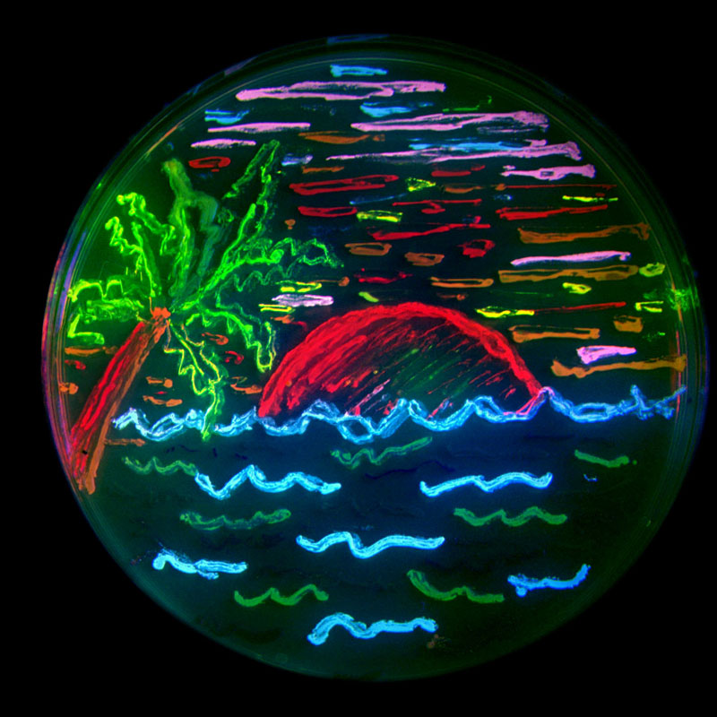
Panel on top shows the fluorescent protein 'palette' developed by Tsien lab - note range of colors and the fruity names. On the left, artwork with bacteria expressing various colors of fluorescent protein.
The major advantage of GFP is that inside a living cell, it can emit light on its own without the help of another protein or other chemicals. It is also possible by using molecular biology techniques, to attach the DNA of GFP to the DNA of the protein of your choice to produce a recombinant DNA. When the information from such recombinant DNA gets translated into a protein within the cell, a tandem protein is created with the GFP unit hanging from the protein. Importantly, since the size of GFP is relatively small, in most cases it does not interfere with the regular functions of the protein it is attached to.
In the simplest of applications, after shining light on the cells, the total amount of fluorescence obtained from the cells provides a measure of the level of expression of the protein tagged with GFP. However the more useful applications involve cells placed under microscopes with high magnifying power (40x to 100x) in conjunction with either arc lamps or lasers for
capturing images of the emitted light. In these cases, we can literally see where the protein of interest is located, or illuminate a particular subcellular structure. For example, the figure on the right shows the mesh of protein network that act as a 'skeleton' (in fact it is called the 'cytoskeleton') in majority of cells in higher organisms. A protein called 'actin' that is involved in this scaffold has been tagged with GFP.
It is also possible to tag two or more proteins in the cells with different fluorescent colors (see the fluorescent protein palette above) and follow their localization or movement in cells. This helps in noting where two proteins are localized 
(in a future post, I will talk about a technique involving fluorescent proteins of two colors which is used to determine if two proteins interact with each other inside a cell)
Perhaps the most powerful application of fluorescent proteins is when you combine microscopy with time lapse video images. In such cases, it is possible to observe where and when the translocation of the protein in cells is taking place under a biological condition. For example, see a video here of a cell moving around with a protein involved in the focal adhesion tagged with GFP.
Visualization of protein location and dynamics in this manner enable scientists to place cells under various physiological conditions and observe the resultant phenotype of the protein behavior. Before the advent of GFP, scientist had to destroy the cells and use other tedious biochemical techniques to obtain similar information. Even then real-time data acquisition was not possible.
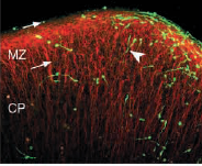
A quick search of the database will reveal more than ten of thousands of peer-reviewed publications where fluorescent proteins have been used to study protein functions at the cellular level. In most of these cases, research was conducted with either unicellular organisms or cells derived from tissues of mammals. However, apart from single cells, fluorescent proteins are also being used at the tissue and even the whole organism level. The picture below shows an example of a research which is investigating the movement of neurons (labeled with GFP) in the cerebral cortex.
A more well-known example of GFP in whole organisms, is the development of 'fluorescent mice' by the company Anticancer Inc . It is easy to follow tumor progression and cancer metastasis in such mice. Also, a Taiwanse reasearch group recently created 'fluorescent pigs'. Stem cells or organs from these pigs when transplanted into other organisms can be followed easily without requiring invasive techniques.


Animal Farm: fluorescent pigs and mice.
More examples of such applications of GFP can be found here. Apart from these animals, 'Alba' , the fluorescent rabbit and fluorescent aquarium fishes are two examples of more esoteric application of this scientific technology.
On a final note, betting markets for the Nobel Prize (yes they do exist !), were predicting this year’s Chemistry Nobel to go to Roger Tsein and others for their work on fluorescent proteins. It eventually went to Roger Kornberg for his work on DNA transcription. Considering the importance of fluorescence proteins and their wide-ranging revolutionary impact on biology, it is not far-fetched to think that the Nobel is not beyond the grasp of these researchers.
Notes:
1. Aequorin itself has been very useful for visualizing cellular calcium concentrations, the regulation of which is important for a number of physiological activities.
2. Without going into great details about physi-chemical mechanisms of GFP fluorescence, suffice to say that the protein has a barrel-like structure (see below); within the barrel, three critical amino acids are brought together in close spatial proximity, which forms the chromophore.
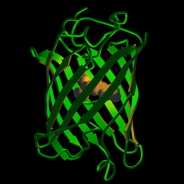
Artistic rendition of the three-dimensional structure of GFP.
3. All images have been linked back to their original web-page.
4. Recommended further reading: This web-site is a very good resource for learning more about GFP's discovery, structure and applications. Also read this interview with Dr. Martin Chalfie.
Coming up: "Much to fret about ": on a technique known as fluorescence resonance energy transfer that enables biological distance measurements, detection of protein interactions, and can be used to look at protein functions at a single molecule level !
Wednesday, October 25, 2006
A Splicing Primer
The central dogma of molecular biology DNA-> RNA-> Protein shows the direction of flow of information of how the cells use the information stored in our DNA to make the necessary proteins. But the situation in most eukaryotes is a little more complex than that simple statement. In most eukaryotes, a gene sequence in a DNA is interrupted by non- coding information. Hence to make a protein, a cell first has to transcribe the gene (make a RNA copy of the gene, called pre-mRNA) and then modify the pre-mRNA by removing the non-coding sequence (intron) and joining the coding sequences (exons) together. The modified mRNA is then exported from the nucleus (where it was made) to the cytoplasm where the ribosome uses it as a template to make the protein. In simple English, the gene for making a proteinA looks like this "HEREabhjhdyfrhUSEndcbldfhdfmMEd ldshhglgmcFORdbfhdflhfnmc PROTEIN A". The task of the cells is to remove the gibberish and make a readable text out of the given instruction - HERE USE ME FOR PROTEINA. The cells then send this information to the ribosome (the protein factory) to make the protein.
Pre-mRNA splicing is the process in which the intronic sequences are removed within a large RNA-protein complex called spliceosome.
Why is splicing important? A spliceosme can remove the non-coding introns present in a given transcript varying combination in response to cellular cues, a process called alternative splicing. The recent completion of a draft of the human genome indicated that more than 59% of the human genes seem to be alternatively spliced (Hastings and Krainer,2001) and thus we can have more complexity (make a larger number of proteins) without increasing the number of genes present. For eg, the Dscam gene in flies has 38,000 alternatively spliced isoforms from four variable exon clusters!
More importantly, it is estimated that aberrant splicing causes about 15% of genetic diseases in humans (Philips and Cooper,2000). Thus, the spliceosome plays a critical role in generating the right template for making a protein and any abnormality in this process would be deleterious to the organism.
What do we know about this process? From genetic and biochemical experiments in the humble budding yeast, scientist have been able to understand how this process occurs. Because both the mechanism of splicing and the splicing machinery are highly conserved throughout eukaryotes, knowledge of yeast splicing gives us insights into the basic process in humans.
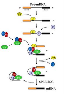 The spliceosome is the largest structure in the cell and is composed of five small nuclear RNAs ( called U1, U2, U4, U5 and U6 snRNAs) and over 100 different proteins (Stevens and Abelson , 2002). Under standard in vitro (i.e. in a test tube) assay conditions, the spliceosome assembles in a step wise manner through the addition of the U1 -> U2-> U4/U6.U5 snRNP particles (the small nuclear RNA along with its associated proteins, represented by a colored blob in the picture) on the pre-mRNA (See Figure). This assembly is an expensive process for the cell as each step consumes energy. But it also allows the apparatus to check each step and hence allows for a greater control over the overall process. Remember, a single mistake here would result in a protein that either does not function or functions abnormally. That to a cell would be hazardous and hence the cells err on the side of caution. After the assembly of the spliceosome, it undergoes structural rearrangements, resulting in the loss of U1 and U4 snRNAs, to become catalytically active (Brow D. A, 2002). Then, it proceeds to remove the intron by two transesterification reactions.
 The resultant message is released from the spliceosome along with the intron. The spliced RNA is exported to the cytoplasm for translation into the protein and the intron degraded by enzymes in the cell. The spliceosome is disassembled and the components (proteins and the snRNAs) recycled for another round of splicing.
Though much is known about the overall process, there is no insights into what triggers the activation. What informs the spliceosome that everything is set in place and hence go ahead and splice? How does the cell control the ATP driven helicases that remodel the spliceosome at each step? Or what cues the cell about abnormal spliceosome and how does it take a stalled spliceosome apart?
Next time I will try and address the role splicing plays in Humans. How does a cell choose which exon to keep? How do DNA elements present in the gene (ISEs) affect choice of exon? Does the rate at which the transcript is made affect exon choice? So keep your eyes out for Splicing -part deux.
References -
Brow D. A, Annu Rev Genet., 2002, Jun 11; 36:333-60.
Hastings and Krainer, Curr Opin Cell Biol., 2001, Jun; 13(3):302-9
Philips and Cooper, Cell Mol Life Sci., 2000, Feb;57(2):235-49
Stevens and Abelson , Methods Enzymol. 2002;351:200-20.
Check this Animation
Saturday, October 14, 2006
A Beautiful Mind
"Imagine if you'd suddenly learnt that the people, the places, the moments most important to you were not gone, not dead, but worse- had never been. What kind of hell would that be?". - A Beautiful Mind, 2001.
I saw the movie for the second time last night and it got me thinking about the complex disorder that is schizophrenia, and the intense effects it has on an individual, making him lose the distinction between real and imaginary. So what is it that makes a person harbour irrational thoughts and so convinced about the his false fears? I tried to poke around the literature to try to understand how much of the organic basis for this disorder is understood. There is the genetic component- the heritable nature of this disorder has been well documented over the years. Mutations in genes that are involved in brain function can be inherited, causing offspring of schizophrenics to be that much more at risk of developing the disorder. The environment plays an equal role, stress and psychological trauma are known to have a causal or triggering effect in schizophrenia, translating genetic predisposition to development of the disorder.
The neuropathology of the disease itself is closely linked to the above described factors. Bad genes, as well as early trauma to the brain, prenatal exposure to infections and psychological trauma result in brain abnormalities that cause cognitive defects and result in the disorder. There are two aspects to understanding how impaired brain function leads to this condition. Firstly the anatomic location of neural systems that are disrupted govern the types of symptoms exhibited by a patient. Various regions of the brain are involved in different functions such as processing impulses, perceiving thoughts and producing a reaction to a stimulus. The distortion in reality observed in schizophrenics is attributed to one region of the brain, thought disorganisation involves malfunction of a different circuit, while a decline in perceptive and physical responses are traced to malfunction in yet another circuit.
Secondly, brain chemistry- in terms of fluctuations in neurotransmitters (the chemicals that transmit signals in the brain cells) control the duration of above mentioned symptoms, to add another layer of complexity to this intricate orchestration. The sum effect of all of this is disorganised thinking, delusional and paranoid thought processes and auditory hallucinations that manifest as schizophrenia.
Dopamine, glutamate and NMDA are some of the neurotransmitters that have been implicated in schizophrenia. The "Dopamine hypothesis" is particularly famous, as it was one of the first major biological causes that could be attributed to schizophrenia. However, it is now thought to be an oversimplification at understanding the disorder, since there are other factors that play a role. Nevertheless, I will discuss the hypothesis because it provides atleast some insight into the process, and is quite fascinating.
Dopamine is a neurotransmitter, and in one of it's functions it is associated with the "pleasure system" of the brain, providing feelings of enjoyment and motivating a person proactively to perform certain activities. Essentially, it mediates the conversion of an outside stimulus from being a "cold" or neutral bit of sensory information into an "attractive" or an "aversive" entity.
For example- normally an external stimulus such as a bright red sports car zipping past a pedestrian might result in a surge of dopamine to cause an appropriate reaction- like the pedestrian turning his head to look at the car. However, the reaction elicited also depends upon the the pedestrian's predispositions and experiences. A race-car enthusiast may turn to look, while a person not interested in sports cars will not exhibit any reaction. In any case, dopamine here mediates a contextually relevant reaction.
In the 1970s, it was discovered that drugs that block dopamine function reduced psychotic symptoms. Further studies led to the hypotheses that dysregulated dopamine transmission causes an abnormal release of dopamine, so that what would have been a normally neutral stimulus results in firing up of neurones and causes aberrant reactions to external objects or their internal representations. Remember Nash in the movie reacting to something as simple as his wife turning on the light by saying "Why did you turn on the lights? Why would you do that? Why?" ?
In this stage, the patient develops a sense of anxiety and confusion, and an intense need to make sense of the new "realities" being experienced. Any and every normal occurrence can produce an exaggerated response in his mind, and he keeps looking for meanings and explanations to calm himself down. As he forms delusions in his mind to explain the occurrences, he experiences a feeling of relief and reduced perplexity. These delusions then persist, even after the stimulus is taken away, eventually taking on a life of their own. Hallucinations arise from similar aberrant thought processes, as the patient conceives an incorrect internal image of a thought or a memory that is percieved and reinforced with such intensity as though it were real.
This is, like I said earlier, just one aspect of cause and development of psychoses but enough to give us a peek into the on-goings in the brain of a schizophrenic. Imbalances in other neurotransmitters and pathways have different ways of interfering with normal thought process and causing psychological disturbances. Given the limited knowledge and understanding of this disorder, how is it brought under control?
Anti-psychotics are useful in the treatment of psychoses, because, in one way, they dampen the effect of the excessive dopamine (in this example) and thus restore a chemical balance that calms the patient. However, they do not change the underlying thought process- all they can do is prevent neutral stimuli from producing abnormal reactions, and quenching aberrant reactions produced initially. Thus, patients are able to "ignore" or control their reactions to stimuli, but are not entirely free of the delusional thoughts that have already formed. This underscores the importance of staying on the drugs as long as is necessary, and also protecting the patient from high stress environments that can cause a resurgence of symptoms. Modern drugs are now being developed to limit side-effects in patients. Imaging technologies have improved to better visualise brain abnormalities associated with schizophrenia. With the availability of genome sequences and better tools, more genes are being discovered that may play a role in the disorder. Emerging tools in pharmacogenomics can make the best of these discoveries to improve treatment. Social acceptance and sensitivity towards the ailment is also needed , to create a support system that does not stigmatise patients.
John Nash's story is a very encouraging one in the face of this complex disorder. His story shows that one can be successful in bringing the disorder under control to a large extent. Eventually, Nash learns to ignore his irrational fears and focus on his passion. The same brain that gave rise to abnormal thought processes also contributed to his Nobel-prize-winning work on the game theory. Indeed, the mind is a beautiful thing!
References: 1) Wikipedia
2) Schizophrenia: challenging the orthodox McDonald et al
3) Schizophrenia in a molecular age. Carol A Tamminga
Monday, October 09, 2006
How does a digital camera work?
In the previous post, I had talked about how a digital photograph is stored in the computer. In this post, I will talk about how a digital photograph senses the photograph talking only about the essential components (using description for RGB colors). A modern digital camera has far more advances than the simplistic picture explained here.
A digital camera has a number of lenses which focus light onto chips that are sensitive to incoming light. In the market, there are two types of image sensors - charge-coupled device (CCD), and the Complementary Metal Oxide Semiconductor (CMOS). CCDs [1] are far more popular than CMOS chips because they are considered to be affected by noise to a lesser extent (and I will use CCDs to explain how a digital camera works). The role of this chip is to sense the light that comes in and convert the light energy to an electric signal that is amplified and then digitized and finally processed.
How a CCD works? Photoelectric effect [2] is the property by which some metals emit electrons when light shines on them. The CCD in the digital camera is a silicon chip that is covered with a grid of small electrodes called photosites. One photosite corresponds to each pixel.
Before a photo is taken, the camera charges the surface of each photosite with electrons. When light strikes a particular side of the photosite, the metal at that site releases some electrons, which travel to the opposite end of the site (forming what is commonly called the capacitor). The larger the intensity of the light that falls on it, the larger the number of electrons that are released, and hence larger the voltage that develops across the photosite. The voltage is then converted to a number using an analog-to-digital converter that corresponds to the intensity of the light that falls on that site. This takes care of the intensity, but we have not discussed about how the photosite knows the color of the light.
As discussed earlier, the color of a pixel is formed by mixing red, green, and blue colors (RGB). So all the light does not hit each photosite, but rather, there is a filter placed on top of the photosite that only lets red, green, or blue color through. Hence, depending on what color is through, each photosite only measures the intensity of the red, green, or blue color that falls on it, and no other color. After this, to measure the intensity of green and blue colors on a site with a red filter, an interpolation algorithm (a process called demosaicing) is used that approximates the intensity of the blue and green light on that site using the intensity of these colors in the neighboring sites.
Lastly, as green is in the center of the spectrum in the visible light (VIBGYOR), our eye is better at picking up different shades of green, and hence, there are a larger number of photosites that sense green light than blue or red. The Bayer pattern shown below is the most common arrangement of photosites in a single array CCD chip.

The other end of expensive digital cameras (read 10's of thousands of dollars) have multiple arrays and avoid the interpolation step. So the incoming light could be split into three copies and then passed through three separate filters and three different arrays and sensed separately to make the final picture by merging these readings together.
There are more complications that arise even in the simple camera, but maybe another post to deal with them (but no promises as I got to do some research before I can post myself).
[1] CCDs were invented by George Smith and Willard Boyle at the Bell Labs.
[2] Albert Einstein won the Nobel prize in 1921 for the quantum explanation of photoelectric effect.
Source:
The source for most of this stuff is Chapter 2 of the Third Edition of the Complete Digital Photography by Ben Long, though the mistakes here are probably mine.
For further reading:
How Stuff Works answers how a camera works
How an image sensor works?
CCD vs CMOS
Wikipedia's CCD entry
A digital camera has a number of lenses which focus light onto chips that are sensitive to incoming light. In the market, there are two types of image sensors - charge-coupled device (CCD), and the Complementary Metal Oxide Semiconductor (CMOS). CCDs [1] are far more popular than CMOS chips because they are considered to be affected by noise to a lesser extent (and I will use CCDs to explain how a digital camera works). The role of this chip is to sense the light that comes in and convert the light energy to an electric signal that is amplified and then digitized and finally processed.
How a CCD works? Photoelectric effect [2] is the property by which some metals emit electrons when light shines on them. The CCD in the digital camera is a silicon chip that is covered with a grid of small electrodes called photosites. One photosite corresponds to each pixel.
Before a photo is taken, the camera charges the surface of each photosite with electrons. When light strikes a particular side of the photosite, the metal at that site releases some electrons, which travel to the opposite end of the site (forming what is commonly called the capacitor). The larger the intensity of the light that falls on it, the larger the number of electrons that are released, and hence larger the voltage that develops across the photosite. The voltage is then converted to a number using an analog-to-digital converter that corresponds to the intensity of the light that falls on that site. This takes care of the intensity, but we have not discussed about how the photosite knows the color of the light.
As discussed earlier, the color of a pixel is formed by mixing red, green, and blue colors (RGB). So all the light does not hit each photosite, but rather, there is a filter placed on top of the photosite that only lets red, green, or blue color through. Hence, depending on what color is through, each photosite only measures the intensity of the red, green, or blue color that falls on it, and no other color. After this, to measure the intensity of green and blue colors on a site with a red filter, an interpolation algorithm (a process called demosaicing) is used that approximates the intensity of the blue and green light on that site using the intensity of these colors in the neighboring sites.
Lastly, as green is in the center of the spectrum in the visible light (VIBGYOR), our eye is better at picking up different shades of green, and hence, there are a larger number of photosites that sense green light than blue or red. The Bayer pattern shown below is the most common arrangement of photosites in a single array CCD chip.
The other end of expensive digital cameras (read 10's of thousands of dollars) have multiple arrays and avoid the interpolation step. So the incoming light could be split into three copies and then passed through three separate filters and three different arrays and sensed separately to make the final picture by merging these readings together.
There are more complications that arise even in the simple camera, but maybe another post to deal with them (but no promises as I got to do some research before I can post myself).
[1] CCDs were invented by George Smith and Willard Boyle at the Bell Labs.
[2] Albert Einstein won the Nobel prize in 1921 for the quantum explanation of photoelectric effect.
Source:
The source for most of this stuff is Chapter 2 of the Third Edition of the Complete Digital Photography by Ben Long, though the mistakes here are probably mine.
For further reading:
How Stuff Works answers how a camera works
How an image sensor works?
CCD vs CMOS
Wikipedia's CCD entry
Friday, September 29, 2006
How is a digital photo stored?
Just as the word digital suggests, a digital camera digitizes every image that it captures. What this means is that a typical rectangular photo is divided into a large number of squares which form the basic picture element called the pixel. Within a given pixel, the color of the image does not change. In the simple case where the pixel can either be black or white, the state of the pixel can be binary - either 0 (white) or 1 (black). The state of each pixel is stored at each location and when all the pixels are put together, you get your image back. Hence, the image is said to be digitized now.
In the real world, most pictures are stored either in color or in various layers of grey scale. For example, when each pixel element is stored as 8 bits, the number of states would be 256 - a number from 0 to 255. 0 represents white, 1 to 254 represents various scales of grey in increasing intensity, and 255 represents black. So when a color photo taken with a digital camera is converted to black and white, the correct term is actually a grey scale photo, because it has varying levels of black and white.
In color photographs, each pixel is made by combining various levels of primary colors. When an artist mixes colors with paints, he combines 2 colors to make a 3rd color. The primary colors are the colors which can combine to make all the colors required in a digital camera. In a typical camera, the primary colors used are Red, Green, and Blue (forming the RGB colors). Another set of primary colors used often are Cyan, Magenta, Yellow, and the Black (called the CMYK colors). Each color set has it's own advantages as it can capture a certain range of colors well. The rest of the post will be based on a RGB photo though it will equally apply to CMYK or any color set.
In the case of a RGB photo, each pixel uses up three storage units (for each color) of certain bit size associated with it. In the case of the 8-bit size storage unit, each pixel will have one storage unit each for intensity of red color (numbers 0 to 255 signifying varying levels of red color similar to the grey scale pictures discussed above), green color, and blue color. The various levels of red, green, and blue colors are mixed to form the actual color of that pixel. A picture stored in this manner would be 24-bit picture because each pixel would be stored in a 24-bit memory unit. The fact that the color of a pixel does not vary can be seen when you blow a picture beyond 100%. When the picture on the left is blown up to look at each pixel, the picture will look as the one on the right:
(Picture courtesy http://photo.net/equipment/digital/basics/)
The larger the resolution of the picture, the larger the number of pixels per inch (PPI). If one were to print a photo on paper, the resolution should be greater than 200 PPI on each side of the rectangle. A 3 MP camera can hence be used to print out photos of the 4" * 6" prints. The greater the resolution of the camera, the larger the size of the photo that can be printed out from it.
I will follow this post with a post on how the digital camera senses each photograph.
To get more information on this:
Photo.net's tutorial
Cambridge's tutorial
Wednesday, September 27, 2006
Living in a distributed world
In this post I had mentioned how ants work together to find a short way past obstacles, and said that they are not the only living things that work in that way. Engineers, though, have a different way of looking at it than pure scientists. These types of systems are called Distributed Control Systems, meaning that there is no central controlling authority, but instead, each individual follows a set of simple rules and this alone is enough to achieve the system's objectives.
Such a concept may seem alien at first. We humans are used to a hierarchical system of getting things done... in governing citizens, in managing corporations, in controlling manufacturing systems, in practically everything, we have a system of smaller parts reporting to progressively larger parts. But there is evidence to show that simple rules followed by a large number of peers can lead to as complicated a global behaviour as might be required.
I'm going to take an example of a non-living thing this time. The following experiment is called Conway's Game of Life. Consider a two dimensional matrix, in which each cell can have a state of "dead" or "alive". Each cell has eight neighbours; if zero or one of them are alive, the cell dies of loneliness. If two or three neighbours are alive, the cell lives. If four or more neighbours are alive, the cell dies of overpopulation. A dead cell "comes to life" if it has exactly three live neighbours.
Depending upon the initial state, an astounding variety of beautiful and complex patterns have been observed to arise out of these simple rules, including different types of oscillators, explosions, firing guns, even moving spaceships!
 A snapshot of the Game of Life Applet: From http://www.ibiblio.org/lifepatterns/
While I used this only to demonstrate that simple rules can result in complex, self-organizing behaviour in a distributed world, the fact remains that a lot of the "management" in nature occurs this way. It doesn't take an engineer to understand the problems with centralized control in a large hierarchy; if you've tried getting something past the bureaucracy of your university, workplace or government, you know it all too well yourself! But engineers understand how difficult it is to design a distributed control system. It is only in extreme circumstances that they do indeed deploy systems in such a manner - for example, in large sensor networks, where thousands of sensor nodes may be released in forests, underwater, anywhere!
We live in a distributed world, in which the sum of parts is inevitably greater than the whole. Sometimes we scientists get a glimpse of it in our computer simulations, but you only need to look out of your window to understand. When you see a flock of birds flying in formation. When you see a beehive. And once you truly understand it, you will see the world through brand new eyes.
We live in a distributed world, in which the sum of parts is inevitably greater than the whole. Sometimes we scientists get a glimpse of it in our computer simulations, but you only need to look out of your window to understand. When you see a flock of birds flying in formation. When you see a beehive. And once you truly understand it, you will see the world through brand new eyes.
Monday, September 25, 2006
X Chromosome Inactivation

That cute kitten is Copycat or CC – the first cloned cat. She is genetically identical to her “mom” (to the right) but phenotypically different. The reason is because of the peculiar phenomenon called X-chromosome inactivation (XCI) that occurs in all the female cells in mammals.
In mammals sex is determined by differential inheritance of the sex chromosomes. Females are XX while males are XY. To compensate for having an extra dosage of X chromosome genes that might be developmentally fatal, the cells in the female embryo randomly select one X chromosome and shut it off (transcriptionally silence it). This “silent” state is maintained throughout life in the female cell. Since the phenomenon is random (each cell independently decides its fate), mammalian females are essentially mosaics. CC is different from her progenitor because the gene for coat color lies on the X chromosome and is randomly silenced in the clone, giving it it’s unique calico pattern.
The phenomenon is evolutionarily conserved in the mammalian lineage – since there is preliminary evidence to suggest that XCI occurs in Prototherians (egg laying mammals) who arose 200-300 million years ago and also in Metatherains (marsupials). Since X and Y differentiation also occurs in these lineages, it is likely that XCI arose as a way of dosage compensation.
How is it done? The random X chromosome inactivation has been linked to a region on the X chromosome, called Xic (X- inactivation center). What is so unique about this region is that all the genes characterized so far are non- coding genes – they make the RNA but this RNA is never used to make a cellular protein. The ncRNA (non coding RNA) from this region are responsible for counting the number of X chromosomes present in the cell, marking one for silencing and maintaining the silenced state. The most talked about transcript from this region is Xist which is essential for the silencing step.
The mechanism of how the ncRNA form Xist or other mapped locus do the inactivation is not yet known. It is most likely that transcription of these genes help to bring in chromatin remodeling factors (things that signal that the chromosome is silenced). Since Xist transcription would take about 30 minutes, during which the RNA is physically attached to the X chromosome, this explanation seems plausible. The unraveling of the mechanism would help us understand another dosage compensation mechanism (that of imprinting) but that is another blog.
Saturday, September 23, 2006
Learning from the humble ant
The next time you're outdoors and you see a trail of marching ants, try a little experiment. Place an obstruction asymmetrically in their path, which is steep enough that they can't climb over it, and see what happens. At first, they would spread out equally in both directions to rejoin the trail. Over a period of time, you'll see the longer trail thin out and disappear as all the ants start taking the shorter trail.
What's really remarkable about this is that ants cannot see and cannot talk. That is to say, they lack the sensory organs for sight and sound. So how do they do this job of finding a short route around obstacles to their target food source?
The secret, as with many other things observed in nature, is elegant in its simplicity and inventiveness. Ants have a very good sense of smell, and lay out a class of chemicals called Pheromones to mark their trail. Each ant lays out roughly the same amount of pheromone for a particular food source, and the pheromone evaporates at a constant rate. When an ant moves towards a marked food source, it tends to move in a direction in which it senses a relatively higher concentration of pheromone.
That's all. If you're puzzled as to why I cut the explanation short, take a minute to think about how this would work and then read on. Now, when the obstacle is placed in the path, the ants can no longer sense the correct direction to travel, and they choose at random among the available directions. Ultimately, they would rejoin the trail, but each ant has now laid pheromone along the path it has taken. Initially, there is little to choose from between the two options, but over a period of time, the shorter path, simply by virtue of being shorter, would have a greater number of ants traversing the path back and forth. This means that the evaporating pheromone on the shorter route gets regenerated more often than on the longer route, causing more ants to choose that direction over the other. This further increases the disparity in pheromone level between the two choices, until there is insufficient pheromone on the longer trail to tempt even a single ant to go in that direction.
This kind of a process is called Auto-Catalytic, meaning it speeds itself up. Some time around 1990, an Italian scientist called Marco Dorigo got a brainwave on how to use this ant behaviour to solve combinatorial optimization problems, which are notoriously difficult to solve with conventional mathematical techniques.
What he did was to represent an optimization problem as a set of obstacles in the path of an ant trail. The decision of which direction to take at an obstacle represented the decision of what value to assign to a variable. In other words, if a variable can take four different values, it is like placing an obstacle giving an ant four different paths around it. Each variable in the problem is equivalent to an obstacle on the trail. "Pheromone concentrations" are maintained virtually for each decision at each obstacle.
The generation of a solution is done randomly, with the probability of taking a particular decision being proportional to the virtual pheromone concentration. Then, the "goodness" of the solution is quantified by the objective function, and this is analogous to the distance traveled to reach the food source by that route. If it is a poor solution, it is a long route and the pheromone concentrations along that route are increased by a small value. If it is a good solution, the pheromone concentrations are increased by a higher value; and all the time, pheromone is "evaporated" at a constant rate at every decision point.
It worked. In a way similar to how ants find paths, an algorithm known as Ant Colony Optimization (ACO) was developed and refined, and although it does not always find the globally best solution, it tends to find a good solution for even large problems in reasonably quick time.
In spite of all the nature-defying technological marvels scientists and engineers come up with, there seems to be an infinite depth to the workings of nature from which we learn continually. The ant optimization algorithm is but one in a wide class of algorithms under the banner of Swarm Intelligence, for ants, bees, birds and many other social denizens of nature have unique and innovative ways of doing a plethora of tasks. Computer scientists observe, and come up with smarter algorithms. Engineers observe, and come up with better control system schemes.
How wrong we are, in talking about "humble ants". It's we who are the "humble scientists".
Links for further reading:
Marco Dorigo's page on the behaviour of real ants that inspired ACO
Wikipedia
Marco Dorigo's landmark paper on Ant Systems
About us
TGFI has always been fascinated by two areas of science the most, astronomy and biology. Over time, the double helix has won her over and she decided to get a Ph.D in molecular biology way back when she was in high school. Fundamental processes in which genes operate, regulate and interact within a system fascinate her the most, which also happens to be the area in which she is getting her Ph.D. Five years at it and she realises how hard it has become for her to think outside her little world. Which is why she think this collaborative blog is a great idea, will make her get out of her world a bit, and talk about science in layman's words. She hopes to contribute on here once a month.
Sakshi is a grad studnet in UK and work on splicing and spliceosome assembly in the budding yeast. Her project focuses on how a cell targets misformed or locked spliceosome and activates turn over. Her interests in science lies in molecular / genetic underpinnings of life. Though she makes frequent forays into other aspects of biology, she is interested in regulation of gene expression (she believes RNA to be the coolest thing, ever). To keep her sanity in the ever crazed world of an underpaid, over-worked graduate student, she indulges in photography, reading (she prides herself on reading everything she can lay her hands on) and listening to music or watching movies.
Prashanth is an incurable romantic and an incorrigible geek from Chennai who tries to convince himself that they are both desirable qualities. He can be bribed into doing almost anything by lending him a good book. He is currently doing his Ph.D. in Industrial Engineering at the Pennsylvania State University.
Murthy is a grad student (and an engineer by birth) who is interested in many things some of them being: Control systems application to biological systems and control systems in nature(They are more interesting and far more elegant than the crude imitations scientists try to build and feel proud about). Algorithms and coding, Fluid mechanics and ofcourse Biology, specifically immune system of mammals and behavior of microorganisms as influenced by evolution are all part of his scientific interests. He is an Agricultural engineer(Specifically Dairy, food Engineer by training) now working in biofuels and control systems.
Back in high-school, BioPondit was aiming to be an engineer, loved physics, but ended up with a undergraduate degree in chemistry. Continuing in this vein, he trained as a Biophysicist in graduate school and is currently engaged in post-doctoral research in Cell Biology, shedding light on cellular signaling mechanisms. Love of books, cinema and an esoteric taste in music is what drives him - that and an ice-cold martini (stirred not shaken). A desire to communicate the beauty of science is what brings him here.
BaL is a grad student in the Chemistry department at the U of I. Broadly, he works on Computational Biophysics, Bioinformatics, and Evolution and these interdisciplinary fields keep him on his toes most of the time. He is also fascinated by cricket, movies (both Hollywood and Bollywood), and photography. He also tends to listen to a lot of desi music (especially Rehman), and classic rock.
Sonya is and always will be a science dork. Since a very young age, she has been fascinated by animal behavior of all aspects. She has long been amazed with how interactions within species, as well as, across species are affected by evolution and how they often eventually lead to speciation events. Although she believes that studying behavior in a natural environment is important, she is now more interested in how neuronal control (neuromodulators) may affect behavior. It is with this goal in mind, that she is working on her Ph.D in a Neurophysiology lab at University of Kentucky and currently working on understanding physiological mechanisms that may influence behavior in invertebrates. Since she is very engrossed in the early stages of her projects, she does not have things to keep her sane. You may on any given day find her talking to her crayfish..luckily they haven’t started talking back yet.
Curious Cat is a theoretical physicist. Her chosen tools of trade include non equilibrium statistical mechanics (the pen and paper kind) and her focus is the dynamics of fluids and fluid-like systems. She is currently post-doc-ing in the New York and Boston area. But this is just the way she earns her bread so to speak. She likes to read widely outside her area of specialization and likes to reduce all that information into minimum models that are easier to understand but still contains the essential features of the true science of the system. And that is why she is a physicist, becuase it gives her the training to be able to do that. She blogs about this and that at http://virtualcuriosityshop.blogspot.com. You can see the few physics posts she has written so far on this blog as well.
Sunday, September 17, 2006
About this Blog
This blog is an attempt to answer any questions we have in mind such that an high school graduate should be able to understand the fundamentals behind the scientific ideas. It could also serve as a portal for us to broaden our knowledge on a subject. The topics that will be discussed will be from a wide range of topics dealing with basic science, and technology. This blog will hopefully be a learning experience for us and it will also be a medium through which we can share our knowledge with each other and with any person who is interested in these topics.
Subscribe to:
Posts (Atom)
